What Is TMS Neuronavigation? Camera-Based and MRI-Based Targeting Explained
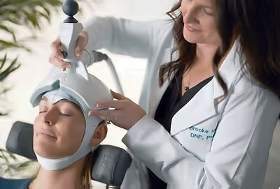
Neuronavigation is the set of technologies used to position a TMS coil at the same spot on a person's head, consistently, from one session to the next. Because a TMS coil's effect is concentrated in a relatively small area of tissue (see how a TMS coil works), even small shifts in coil placement can mean the field is being delivered to a slightly different location than intended. Neuronavigation systems solve this by tracking the coil's position relative to the head in real time, ranging from simple physical landmarks to camera-based tracking to full MRI-based mapping.
This is fundamentally a targeting and workflow question, not a treatment-outcome question—it's about how precisely and repeatably a coil can be returned to the same location, which is an engineering property of the positioning system rather than of the treatment itself. Ampa One includes camera-based neuronavigation as a standard part of its system, at no added hardware cost, which places it at a specific point along a broader spectrum of targeting approaches.
Why does consistent coil positioning matter as an engineering problem?
A TMS coil's magnetic field is strongest in a fairly small zone directly beneath it, and that zone shifts with the coil. If a coil is placed a centimeter or two off from where it was placed in a prior session, the underlying tissue being stimulated changes correspondingly. From a pure positioning-engineering standpoint, this is analogous to any precision-placement problem: the more repeatably a tool can be returned to the same coordinates, the more consistent the physical process being delivered.
Early TMS systems relied entirely on external anatomical landmarks and measurements—for example, measuring distances from the ears or nose to estimate a scalp location, sometimes marked with a cap or grease pencil. This works as a rough approximation but is inherently limited in precision, since it depends on manual measurement and doesn't account for individual variation in head shape or shifts in coil angle during a session.
What are the different TMS positioning approaches?
TMS coil positioning methods generally fall along a spectrum from simple to sophisticated, each with different levels of precision and different equipment requirements.
Landmark-based positioning is the oldest approach: an operator measures reference points on the skull (often based on the international 10-20 system used in EEG placement) and marks or estimates a target location by hand. It requires no additional hardware beyond a measuring tape or cap, but it depends heavily on operator consistency and doesn't adapt to the person's specific anatomy.
Camera-based neuronavigation adds a tracking layer: a camera system monitors the position of the coil and the person's head in real space, typically using reflective markers or facial/head-tracking software, and displays or maintains the coil's position relative to a previously established target. This allows a system to track drift in real time and alert an operator if the coil has moved off target, without requiring an individual MRI scan.
MRI-based neuronavigation goes a step further by using a person's own structural MRI scan to build a 3D model of their head and brain, then registering the coil's position against that specific anatomical model. This allows targeting based on the person's own cortical geometry rather than population-average landmarks or scalp measurements, at the cost of requiring an MRI scan and additional software to register the coil position against it.
How does camera-based tracking work without an individual MRI scan?
Ampa’s camera-based system solves the tracking problem differently than MRI-based systems. Rather than mapping the coil's position against a personalized brain scan, a camera system establishes a reference point on the person's head at the start of a session (or across sessions) and then continuously tracks the coil's position and angle relative to that reference using optical tracking—similar in principle to motion-capture technology used in other fields.
The camera detects markers, distinctive features, or head geometry and calculates, many times per second, whether the coil is still at the intended position and angle. If the coil drifts, the system can flag the deviation so the operator can correct it. This gives a meaningful precision improvement over landmark-and-measurement approaches, without requiring the additional imaging step, hardware, and registration workflow that MRI-based navigation involves.
Because the camera and its tracking software are integrated into the system itself rather than requiring a separate imaging device, this approach can be included as a standard part of a TMS platform. Ampa One's camera-based neuronavigation works this way, tracking coil position throughout a session without requiring separate MRI hardware.
What are the trade-offs in cost and workflow between these approaches?
Each step up the positioning spectrum trades added cost and workflow complexity for added targeting precision, and the right balance depends on the goals of a given system.
Landmark-based positioning has the lowest equipment cost and simplest workflow, but the least built-in consistency check—there's no automated way to detect drift during a session. Camera-based navigation adds real-time tracking and drift detection without requiring individual imaging, which adds some hardware and software but no separate scanning step or per-person imaging cost. MRI-based navigation offers the most anatomically personalized targeting but requires obtaining and processing an individual MRI scan, along with software to register the coil against that scan, which adds both cost and scheduling complexity to the overall workflow.
This is why coil focality (see our comparison of figure-of-8 and H-coil designs) and positioning technology are often discussed together: a highly focal coil benefits more from precise, repeatable positioning, since small placement shifts matter more when the field itself is concentrated in a small area.
Frequently Asked Questions
Is neuronavigation required for TMS to function at all? No. A TMS coil will generate its magnetic field regardless of the positioning method used. Neuronavigation is a workflow and consistency tool, not a requirement for the underlying physics of coil operation.
Does camera-based neuronavigation use radiation or imaging scans? No. Camera-based systems use optical tracking, similar to motion-capture technology, rather than any imaging modality like MRI or CT. No radiation or scanning is involved in the camera-tracking process itself.
How is a target location initially identified before neuronavigation tracks it? Initial target identification typically still relies on established methods, such as anatomical landmarks, standardized head-measurement systems, or (in MRI-based systems) the structural scan itself. Neuronavigation's role is primarily to maintain and verify that position once it's established, not to determine it from scratch.
Does neuronavigation change how the TMS pulse itself works? No. Neuronavigation is a positioning and tracking system layered on top of the coil and stimulator; it doesn't alter the electromagnetic pulse generation described in our article on how a TMS coil works. It affects where the coil is placed, not how the field is generated.
Why does Ampa One include camera-based neuronavigation as standard rather than optional? Camera-based tracking can be built into a system's core hardware and software rather than requiring separate per-session imaging equipment, which is why it can be offered without an added hardware fee. This reflects a design choice about how the positioning system is integrated into the overall platform.
Is MRI-based neuronavigation always more precise than camera-based tracking? MRI-based navigation offers anatomically personalized targeting based on an individual's own brain structure, which is a different kind of precision than real-time position tracking. Camera-based systems excel at detecting drift and maintaining a consistent position throughout a session; MRI-based systems excel at establishing a target based on individual anatomy. The two approaches address related but distinct parts of the targeting problem.
This article is for general educational purposes only and is not medical advice. For questions about how targeting technology applies to a specific treatment plan, consult a qualified clinician.

Meet the TMS system built for what’s next
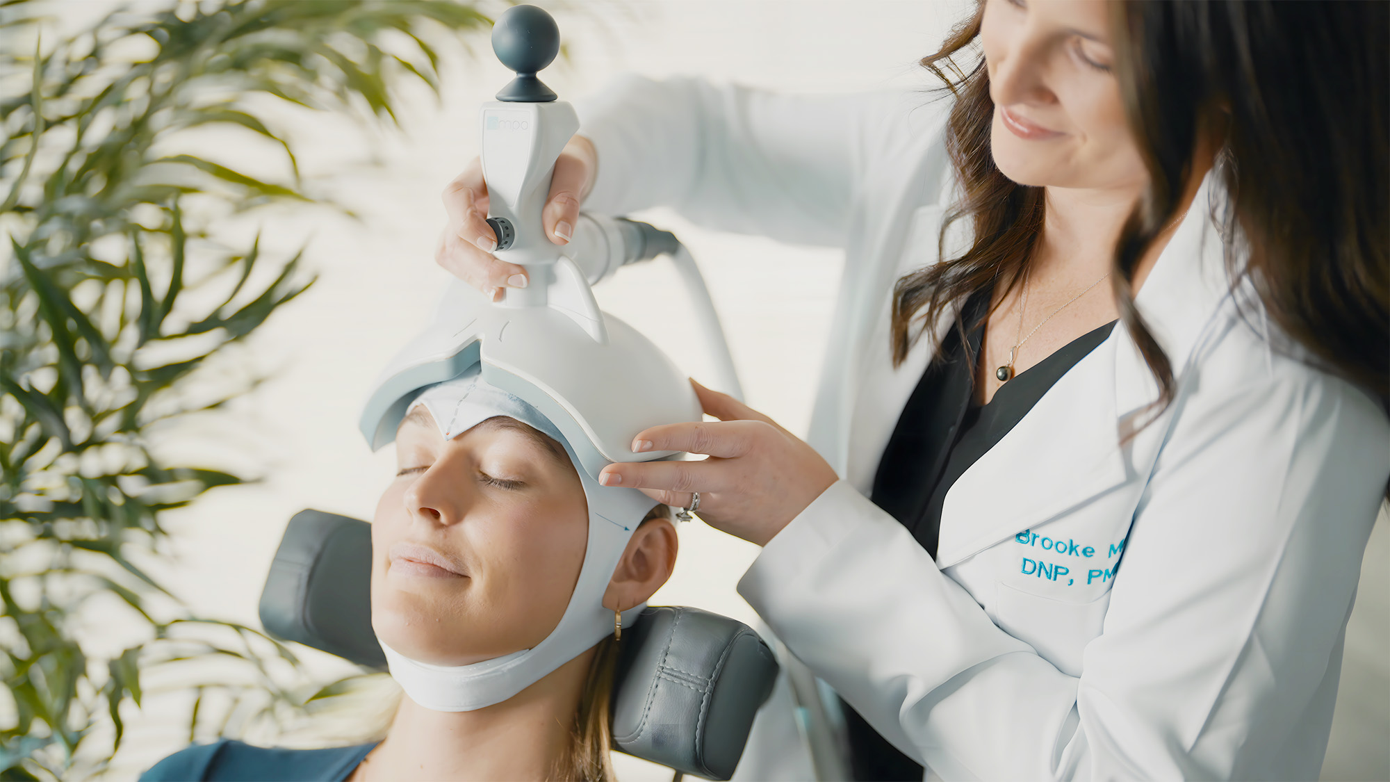
Ampa One brings accelerated protocols, precision neuronavigation, a compact footprint, and a flexible subscription model together in one streamlined TMS platform.
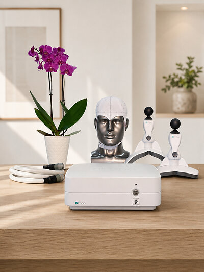
FDA-Cleared TMS Devices Compared: Ampa One, NeuroStar, BrainsWay, MagVenture & Magstim
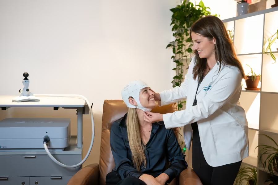
How to Prepare for Your First TMS Appointment: A Practical Checklist